Manual Therapy Online
Technique Peek - Foot and Ankle Manipulations
1. Talocrural Subluxation
This is possibly one of the most common biomechanical dysfunctions in the peripheral system. It is a frequent accompaniment to an inversion injury. Following inversion injury, the talus may become anteriorly fixed. This should mean that it couldn't glide posteriorly at all, therefore completely limiting dorsiflexion of the ankle. However, only a few degrees of dorsiflexion are actually lost. The subluxation therefore cannot be anterior. Rather the talus is subluxed into medial rotation. This would completely limit lateral rotation, which is the conjunct rotation of dorsiflexion. As only the final part of dorsiflexion relies on conjunct rotation, only a few degrees of range are lost. Rarely, a posterior subluxation will be encountered, the traction manipulation is appropriate for this condition but the J-stroke manipulation, because it is directional and posterior is not.
Anatomy and Biomechanics
The ankle is a modified sellar synovial joint with one degree of freedom, plantaflexion and dorsiflexion. The talus presents a convex surface to the cruris for the degree of freedom therefore its arthrokinematic is opposite its osteokinematic. For dorsiflexion it glides posteriorly and for plantaflexion anteriorly, Its stability is dependent on its collateral ligaments and the integrity of the inferior tibiofibular joint, which in turn relies on anterior, posterior and interosseus ligaments. During the last few degrees of dorsiflexion, the talus rotates laterally as a conjunct movement.
Examination Findings
The passive physiological motion of dorsiflexion is limited by 5-10 degrees by an abrupt hard (but not bony) end feel. The same end feel limits the posterior talar glide. Plantaflexion and anterior glide are normal with either a normal capsular or a soft capsular end feel (if the anterior capsule has been over-stretched). If the talar swing test has been utilized in addition to the above, the tester will feel the loss of the lateral rotation of the talus with dorsiflexion.
Technique
 The common treatment for this condition is done in error. The mistake is in believing that the plantaflexors which are tight. This idea can easily be dispelled. First, it cannot be the gastrocnemius as the constant length phenomenon is absent when the knee is flexed. So it could be the soleus, but this would not limit the arthrokinematic (joint glide) nor alter its end feel, both of which are noted on examination. In fact stretching the plantaflexors is exactly the wrong treatment. It will eventually restore the range of dorsiflexion but it usually does so by spreading the mortise formed between the tibia and fibula. Instead of this, a simple manipulation usually effects a cure. The technique that will be described is the traction manipulation. This is non-directional as far as the lesion is concerned.
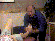
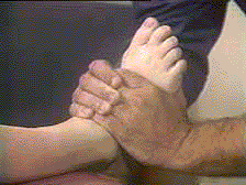
The problem joint is distracted and then let go to reposition normally (with any luck). The patient lies supine with the unaffected leg's knee flexed to protect the back (if back pain is present, the patient can pull the knee up to the chest). The PT stands in line with the leg by the foot. The PT's hands are wrapped around the instep of the foot so that the little or ring finger of one hand is over the neck of talus. The thumbs lay along the sole of the foot and in this position, the thumbs maintain the posture of the foot, preventing it from dorsiflexion when the thrust is applied. The foot is positioned in its resting (neutral) position to permit maximum room within the joint. The PT pulling the hands towards the body takes up partial distraction slack. The manipulation is effected by a sharp pull distally in line with the tibia, causing the talus to distract from the mortise.
 The J-stoke manipulation is reserved for the subluxation resistant to the simple traction technique. This is a combination of distraction and posterior thrust causing a scooping movement to occur (the J). Much care must be taken to ensure that dorsiflexion does not occur during the manipulation.
2. Talocalcaneal Subluxation
 The calcaneus can sublux into inversion or eversion under the talus. Inversion subluxations often result from inversion injuries of the ankle especially where the weight is taken through the heel rather than the cuboid area. Eversion subluxations may result from over-flattening of the foot for whatever reason.
Anatomy and Biomechanics
This is structurally a modified ovoid joint but functionally it is a modified sellar. The reason for this is that the joint is essentially two joints in one. The superior surface of the calcaneus is concave anteriorly while posteriorly it is convex. As both surfaces have to move simultaneously, it means that they effectively form a concavoconvex or sellar surface. Consequently this joint has only one degree of freedom, inversion and eversion. During inversion, the anterior surface being concave moves with the osteokinematic that is medially while the posterior surface moves in the opposite direction, that is laterally.
The collateral ligaments of the ankle, small talocalcaneal ligaments and the two bands of the interosseus ligament afford stability of the joint particularly the posterior.
Examination Findings
Inversion subluxations are characterized by a loss of passive physiological eversion, especially the roll laterally at the end of the movement. The end feel is jammed (i.e. abrupt and hard). The arthrokinematic of posterior talar gliding is also jammed. Eversion subluxations are recognized by limited physiological inversion and anterior talar gliding, again both with a pathomechanical end feel. Either subluxation may occur at the anterior or posterior joint surface with the anterior perhaps being more common. To determine which joint surface is affected, medial and lateral glides are performed across the joint. The posterior calcaneus presents a convex surface to the talus so if this surface is glided laterally, it tests the medial osteokinematic that is inversion and if glided medially, eversion. The anterior joint surface of the calcaneus is concave so the arthrokinematic and osteokinematic are in the same direction. Consequently, a medial glide tests inversion and a lateral eversion.
Technique
Eversion Manipulation (Inversion Subluxation)
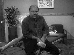
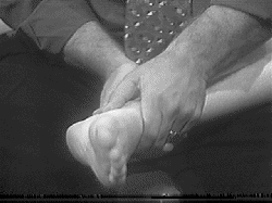
 The patient lies on their side with the medial border of the affected foot upwards. The therapist sits on the bed with his or her back towards the patient. The foot is held so that the thumbs are positioned over the lateral aspect of the calcaneus over either the anterior (sustentaculum tali) or posterior (inferior to the medial talar tubercle) joint line depending on which is dysfunctional.
The adjacent figures show the thumb positions for an eversion manipulation (inversion subluxation) with the thumbs positioned on the sustentaculum tali.
The foot is lifted into eversion and then flicked downwards into eversion with pressure being applied by the thumbs at the end of the flick. This is an osteokinematic manipulation so the direction of the manipulation is always lateral regardless of whether the anterior or posterior joint is involved.
Inversion Manipulation (Eversion Subluxation) To manipulate to gain inversion (an eversion subluxation), the patient lays so that the lateral border of the foot is uppermost. The therapist takes up the same position but now with the other leg. The thumbs are placed over the lateral border of the calcaneus. The posterior joint is found as the area just posterior and inferior to the lateral malleolus and the anterior being just posterior to the cuboid and just below and anterior to the malleolus. Again this is an osteokinematic manipulation so the thrust is always downwards that is into inversion.
Featured Course
FREE COURSE
Foundations of Expertise
Date:
Starting March 24, 2024
Location: Online
LEARN MORE


